Table of Contents
Introduction
The femur bone, often called the thigh bone, is one of the most remarkable structures in the human body. It is the longest, strongest, and heaviest bone we have, capable of withstanding extreme forces and playing a vital role in mobility. From the moment we take our first steps as children to the way we run, jump, and stand in our daily lives, the femur is at the center of our skeletal strength.Anatomically, the femur links the hip joint to the knee joint, bearing the body’s weight while enabling strong and dynamic leg movements. Its unique design makes it not only essential for walking and running but also for absorbing shocks
This comprehensive guide will explore the structure of the femur bone, its functions, common injuries, diagnosis, treatments, prevention, and rehabilitation. By the end, you’ll have a complete understanding of why the femur is such a crucial part of human anatomy and how to protect it for a lifetime of healthy movement.
________________________________________
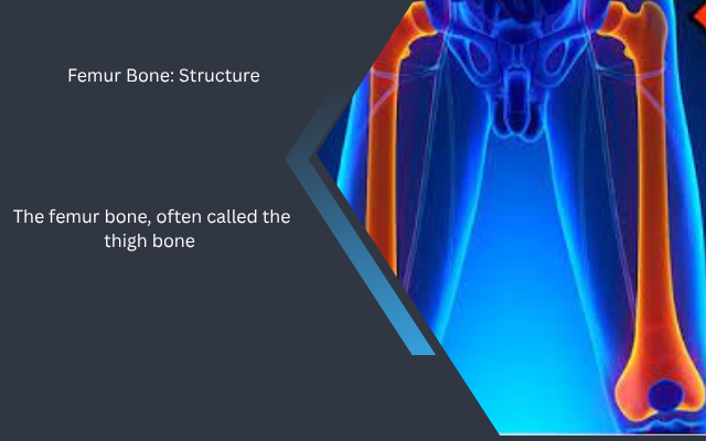
1. Detailed Structure of the Femur Bone
The femur is located in the upper leg, extending from the hip to the knee. It is classified as a long bone and has several distinct features.
1.1 Main Parts of the Femur
1. Proximal End (Upper End)
o Head: The rounded ball that fits into the hip socket (acetabulum), forming the hip joint.
o Neck: A narrow section connecting the head to the shaft, common site for fractures in older adults.
o Greater Trochanter: A large, bony prominence on the outer side of the femur where muscles attach.
o Lesser Trochanter: A smaller bump located just below the neck, also serving as a muscle attachment point.
2. Shaft (Body of the Femur)
o Strong, cylindrical section that bears weight.
o Encased in a dense layer of compact bone that provides exceptional strength
o Contains the medullary cavity, filled with bone marrow.
3. Distal End (Lower End)
o Medial and Lateral Condyles: Rounded areas that articulate with the tibia to form the knee joint.
o Patellar Surface: Groove where the kneecap (patella) rests.
________________________________________
1.2 Bone Composition
The femur is made up of:
• Compact Bone: Dense outer layer providing strength.
• Spongy Bone (Cancellous Bone): Lighter, porous inner structure found near the ends of the bone.
• Bone Marrow: Produces red blood cells, white blood cells, and platelets.
________________________________________
1.3 Blood Supply and Nerves
• The femoral artery provides oxygen-rich blood to the bone.
• Femoral nerve and other nerve branches supply sensation and motor control to surrounding muscles.
• Healthy blood flow is critical for bone healing after injury.
________________________________________
2. Functions of the Femur Bone
The femur’s primary role is structural support, but its importance extends much further.
2.1 Weight Bearing
• Supports the body when standing, walking, and running.
• Distributes weight evenly to prevent strain on other bones.
2.2 Movement
• Functions in coordination with the hip and knee joints to enable leg movement
• Allows flexion, extension, abduction, adduction, and rotation.
2.3 Shock Absorption
• Cushions the body from high-impact activities such as jumping or sudden stops.
2.4 Muscle Attachment
• Serves as an anchor for major muscle groups like the quadriceps, hamstrings, and gluteals.
________________________________________
3. Common Injuries to the Femur Bone
Despite its strength, the femur can be injured. Such injuries are often severe and demand immediate medical intervention.
3.1 Femur Fracture
• Cause: High-impact trauma from car accidents, falls, or sports.
• Symptoms: Severe pain, inability to stand, swelling, deformity.
• Treatment typically involves surgical intervention using metal rods, plates, or screws for stabilization..
________________________________________
3.2 Stress Fracture
• Cause: Often results from repetitive stress, frequently seen in runners and military personnel
• Symptoms: Gradual pain during activity, tenderness along the thigh.
• Treatment: Involves rest, a gradual return to physical activity, and occasionally the use of a walking boot for support.”
________________________________________
3.3 Hip Fracture (Femoral Neck)
• Cause: Often from falls in elderly patients with osteoporosis.
• Symptoms: Groin pain, inability to walk, leg shortening.
• Treatment: Surgical repair or hip replacement.
________________________________________
3.4 Open Fractures
• Bone breaks through the skin.
• High risk of infection, requiring emergency treatment.
________________________________________
4. Diagnosis of Femur Injuries
• Physical Examination: Checking range of motion, swelling, tenderness.
• X-Rays: Standard tool for fracture detection.
• MRI or CT Scan: Used for complex fractures or soft tissue damage.
________________________________________
5. Treatment Options
5.1 Non-Surgical Treatment
• Suitable for minor stress fractures.
• Includes rest, pain management, and physical therapy.
5.2 Surgical Treatment
• Intramedullary Nailing: Rod inserted into the bone for stability.
• Plates and Screws: Used when fracture is near the joint.
• Hip Replacement: For severe femoral neck fractures.
________________________________________
6. Rehabilitation and Recovery
6.1 Recovery Phases
1. Early Phase: Pain control, limited movement.
2. Strengthening Phase: Gradual exercises to restore strength.
3. Return to Activity: Progressive loading and functional movements.
6.2 Recovery Time
• Simple fractures: 3–4 months.
• Severe injuries: 6–12 months.
________________________________________
7. Complications
• Nonunion: Bone fails to heal.
• Malunion: Bone heals incorrectly.
• Infection: Especially in open fractures.
• Blood Clots: Risk increases after surgery.
________________________________________
8. Prevention
• Strengthen leg muscles.
• Maintain bone density with calcium and vitamin D.
• Use protective sports gear.
• Prevent falls with balance exercises.
________________________________________
9. Interesting Facts
• The femur is about 26% of a person’s height.
• The femur is capable of withstanding forces up to 30 times a person’s body weight
• Archaeologists use femur measurements to estimate height.
________________________________________
10. Frequently Asked Questions (FAQs)
1. What is the typical healing time for a fractured femur?
Recovery can take 3–12 months depending on severity.
2. Can you walk with a broken femur?
No, a fractured femur usually makes walking impossible until treated.
3. What foods help strengthen the femur?
Calcium-rich foods like dairy, leafy greens, and fortified cereals; plus vitamin D sources like fish and sunlight.
4. Are femur fractures common in children?
They are less common in children due to more flexible bones but can still occur from high-impact falls. ________________________________________
Conclusion
The femur bone is a vital part of your body’s mobility system. Understanding its structure, function, and potential injuries helps in prevention and recovery. With the right care, nutrition, and exercise, you can maintain strong and healthy bones for life.